Article Type: Short Report, Volume 1 Issue 1
*Corresponding author: V Lunde Dadon
Department of Osteopathic Robotics Surgery, Clincial Research Board of Governing Didactic Research Series, Outlying Islands.
Email: mllcsage808@gmail.com
Received: Nov 15, 2024 Accepted: Dec 04, 2024 Published: Dec 11, 2024
Citation: Dadon VL, Dadiane L. Interventional pillars in robotics and didactics approach to imaging modalities: Radiology and robotics in 2025. Ann Case Rep Med Images. 2024; 1(1): 1011.
Copyright: Lunde Dadon V et al. © All rights are reserved
In this brief review we will discuss the automation of muscoskeletal functions in nerve signals, and motions derived from osteopathic medicine. We will them merge this knowledge with the new approach to robotics and didactics in Interventional radiology. Briefly we will discuss in this series the current and future applications of robotics into medical Imaging, and natural sciences of muscoskeletal functions. The short summary will discuss one case involving cartilage, bone, and robotic interventional radiology.
Keywords: Didactics; Osteopathic images; Bone and cartilage; Education; Bio-Rad; Bio-Med; Research; Muscokeleta; Computer applications; Technology and robotics in medicine.
1. In the applications of Medical Images we discussed that a brief summary of didactics and robotics was applied to a specimen with cartilage and bone dysfunction.
2. The orbital portion of the femur had a cartilage to bone injury, in this case we manipulated the motion, and soft tissue through invasive robotics, through didactics approach.
3. The image-guidance were analyzed and the percutaneous and muscoskeletal features were technically emphasized in the slides by the Robotics approach.
4. We then navigated through didactic tools, and using the robotic guidance system created a potential to simplify the procedure and targeted the approach to the anatomy by using Medical Images such as CT (Computed Tomography, fluoroscopy, and Ultrasound (US).
1. Scenario-Based results of this medical imaging series case pointed out the osteochondritis in the IR imaging the bone fragments, and cartilage needed a robotic guided approach to separate the conjoined anamoly.
2. We proceeded to contribute to the results for further research into the bone density, size, and percutaneous imaging with the robotic and didactic efforts.
3. The diagnosis wasn’t consolidated, due to intervention that is further required when working with osteopathic features and we issued a CT-Scan and Xray.
4. Upon receiving results, we completed our introductory studies with the medical imaging and IR, and used computer data science to set data points in didactic robotic guidance imaging, and STEM cell research into bone, cartilage, and anamolies, we also identified the osteochondritis and developed a new approach to muscoskeletal manipulation to separate the bone from cartilage.
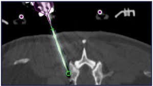
Figure 1: Example of robotic arm guided IR-Into bone/cartilage osteochondritis approach.
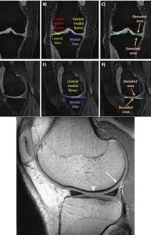
Figure 2: Examples of didactics in identification of needle guided_IR approach (Femur/Bone/Cartilage) in image above.
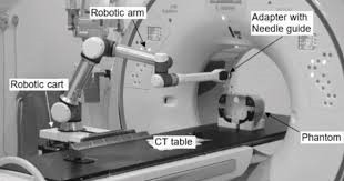
Figure 3: IR-Clinical application procedure room with compliant identification of didactics for robotic interventional imaging, and guided IR-Surgical preparation.
1. In this new applied approach, we remarkable proposed the case to undergo further research.
2. The cartilage and bone were remarkable seperated by IR Guided Didactics, and the Medical Images created in formatics systems in synchronicity with our Research and Development
3. We captured several regenerative features, and specimens for further STEM Cell, and osteopathic case research, and for solutions to IR Guided Didactic Robotics Medical Images.
Author contributions: Governing Board of Didactic and Robotic Research, Non-Profit Facility and Clinicals for Post Doctor ate Students.
Funding: FFIEC, MDPI
Conflict of interests: The authors declare conflict of interest in areas of North America, and Europe. Laboratory location remain soveirgn, and privately funded since 2009.